Cellular tissue
Tissue is a cellular organizational level intermediate between cells and a complete organism. A tissue is an ensemble of similar cells and from the same origin, that together carry out a specific function. These are called tissues because of their identical functioning. Organs are then formed by the functional grouping together of multiple tissues.
The study of tissue is known as histology or, in connection with disease, histopathology. The classical tools for studying tissues are the paraffin block in which tissue is embedded and then sectioned, the histological stain, and the optical microscope. In the last couple of decades, developments in electron microscopy, immunofluorescence, and the use of frozen tissue sections have enhanced the detail that can be observed in tissues. With these tools, the classical appearances of tissues can be examined in health and disease, enabling considerable refinement of clinical diagnosis and prognosis.

Animal tissue.
Animal tissues can be grouped into four basic types: connective, muscle, nervous, and epithelial. Multiple tissue types comprise organs and body structures. While all animals can generally be considered to contain the four tissue types, the manifestation of these tissues can differ depending on the type of organism. For example, the origin of the cells comprising a particular tissue type may differ developmentally for different classifications of animals.
The epithelium in all animals is derived from the ectoderm and endoderm with a small contribution from the mesoderm, forming the endothelium, a specialized type of epithelium that comprises the vasculature. By contrast, a true epithelial tissue is present only in a single layer of cells held together via occluding junctions called tight junctions, to create a selectively permeable barrier. This tissue covers all organismal surfaces that come in contact with the external environment such as the skin, the airways, and the digestive tract. It serves functions of protection, secretion, and absorption, and is separated from other tissues below by a basal lamina.
Connective tussue.
Connective tissues are fibrous tissues. They are made up of cells separated by non-living material, which is called extracellular matrix. Connective tissue gives shape to organs and holds them in place. Both blood and bone are examples of connective tissue. As the name implies, connective tissue serves a "connecting" function. It supports and binds other tissues. Unlike epithelial tissue, connective tissue typically has cells scattered throughout an extracellular matrix.

Muscle tissue.
Muscle cells form the active contractile tissue of the body known as muscle tissue or muscular tissue. Muscle tissue functions to produce force and cause motion, either locomotion or movement within internal organs. Muscle tissue is separated into three distinct categories: visceral or smooth muscle, which is found in the inner linings of organs; skeletal muscle, in which is found attached to bone providing for gross movement; and cardiac muscle which is found in the heart, allowing it to contract and pump blood throughout an organism.

Nervous tissue.
Cells comprising the central nervous system and peripheral nervous system are classified as neural tissue. In the central nervous system, neural tissue forms the brain and spinal cord and, in the peripheral nervous system forms the cranial nerves and spinal nerves, inclusive of the motor neurons. Nervous tissue functions to transmit messages.
Epithelial tissue.
The epithelial tissues are formed by cells that cover the organ surfaces such as the surface of the skin, the airways, the reproductive tract, and the inner lining of the digestive tract. The cells comprising an epithelial layer are linked via semi-permeable, tight junctions; hence, this tissue provides a barrier between the external environment and the organ it covers. In addition to this protective function, epithelial tissue may also be specialized to function in secretion and absorption. Epithelial tissue helps to protect organisms from microorganisms, injury, and fluid loss. Functions:
- the cell of the body surface form the outer layer of skin.
- inside the body,epithelial cells forms lining of mouth & alimentary canal & protect these organ.
- epithelial tissues help in absorption of water & nutrient.
- epithelial tissues help in elimination of waste product
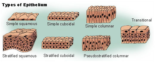
The different types of epithelial tissues are as follows:
- Squamous epithelium,
- Cuboidal epithelium,
- Columnar epithelium,
- Glandular epithelium,
- Ciliated epithelium
Hypoxia.
Hypoxia is a pathological condition in which the body as a whole (generalized hypoxia) or a region of the body (tissue hypoxia) is deprived of adequate oxygen supply. Variations in arterial oxygen concentrations can be part of the normal physiology, for example, during strenuous physical exercise. A mismatch between oxygen supply and its demand at the cellular level may result in a hypoxic condition. Hypoxia in which there is complete deprivation of oxygen supply is referred to asanoxia.
Hypoxia differs from hypoxemia. In the latter, the oxygen concentration within the arterial blood is abnormally low. . It is possible to experience hypoxia and have a low oxygen content (e.g., due to anemia) but maintain high oxygen partial pressure (pO2). Incorrect use of these terms can lead to confusion, especially as hypoxemia is among the causes of hypoxia (in hypoxemic hypoxia).
Generalized hypoxia occurs in healthy people when they ascend to high altitude, where it causes altitude sickness leading to potentially fatal complications: high altitude pulmonary edema (HAPE) and high altitude cerebral edema (HACE). Hypoxia also occurs in healthy individuals when breathing mixtures of gases with a low oxygen content, e.g. while diving underwater especially when using closed-circuit rebreather systems that control the amount of oxygen in the supplied air. A mild and non-damaging intermittent hypoxia is used intentionally during altitude trainings to develop an athletic performance adaptation at both the systemic and cellular level.

Hypoxia Symptoms
The symptoms of generalized hypoxia depend on its severity and acceleration of onset. In the case of altitude sickness, where hypoxia develops gradually, the symptoms include headaches, fatigue, shortness of breath, a feeling of euphoria and nausea.
In severe hypoxia, or hypoxia of very rapid onset, changes in levels of consciousness, seizures, coma, priapism, and death occur. Severe hypoxia induces a blue discolouration of the skin, called cyanosis.
Because hemoglobin is a darker red when it is not bound to oxygen (deoxyhemoglobin), as opposed to the rich red colour that it has when bound to oxygen (oxyhemoglobin), when seen through the skin it has an increased tendency to reflect blue light back to the eye. In cases where the oxygen is displaced by another molecule, such as carbon monoxide, the skin may appear 'cherry red' instead of cyanotic.
Free radicals.
Radicals (often referred to as free radicals) are atoms, molecules, or ions with unpaired electrons or an open shell configuration. Free radicals may have positive, negative, or zero charge. With some exceptions, these unpaired electrons cause radicals to be highly chemically reactive.
Free radicals play an important role in combustion, atmospheric chemistry, polymerization, plasma chemistry, biochemistry, and many other chemical processes. In living organisms, superoxide and nitric oxide and their reaction products regulate many processes, such as control of vascular tone and thus blood pressure. They also play a key role in the intermediary metabolism of various biological compounds. Such radicals can even be messengers in a phenomenon dubbed redox signaling. A radical may be trapped within a solvent cage or be otherwise bound.

No hay comentarios:
Publicar un comentario