Organelles and diseases.
There are two main types or categories of cells: prokaryotic cells and eukaryotic cells. Both of these types of cells have several things in common. All cells are surrounded by a plasma membrane, which is made of a double layer (a bilayer) of phospholipids. Within this membrane, is the cytoplasm which is composed of the fluid and organelles of the cell.
Bacteria (Kingdom Monera) are prokaryotes. They do have DNA, but it is not organized into a true nucleus with a nuclear envelope around it. Also, they lack many other internal organelles such as mitochondria and chloroplasts.
![[Phospholipid Bilayer]](http://biology.clc.uc.edu/graphics/bio104/membrane.jpg)
Organisms in the other four kingdoms are eukaryotes. Their DNA is organized into a true nucleus surrounded by a nuclear envelope which consists of two bilayer membranes. The nucleus of eukaryotic cells contains the genetic material which chemically directs all of the cell’s activities. Usually this is in the form of long strands ofchromatin made of DNA and affiliated proteins. When a cell is getting ready to divide, the chromatin coils and condenses into individual, distinguishable chromosomes. Because the nuclear envelope consists of two bilayer membranes, there is a space between these two membranes called a lumen.
![[Generic Eukaryotic Cell]](http://biology.clc.uc.edu/graphics/bio104/cell.jpg)
Branching off from and continuous with the outer membrane of the nuclear envelope is a double walled space which zigzags through the cytoplasm. This is the endoplasmic reticulum (ER for short) and its central space or lumen is a continuation of the lumen between the membranes of the nuclear envelope. There are two kinds of ER: smooth ER and rough ER. Typically ER closer to the nucleus is rough and that farther away is smooth. Smooth ER is a transition area where chemicals like proteins the cell has manufactured are stored in the lumen for transportation elsewhere in the cell. Pieces of the smooth ER called vesicles pinch off from the smooth ER and travel other places in the cell to transfer their contents. Rough ER gets its name because it has other organelles called ribosomes attached, which give it a rough appearance when viewed by an electron microscope. Rough ER and its associated ribosomes are involved in protein synthesis, with the new polypeptide being threaded into the lumen of the ER as it is formed.
Ribosomes are special organelles that are directly involved in protein synthesis. They are made of RNA (ribonucleic acid) and protein and are manufactured in the nucleus (from a DNA template), then go out into the cytoplasm to function. Ribosomes of prokaryotes and eukaryotes are chemically different enough that some of our antibiotics such as tetracycline, streptomycin, and the new Zithromax® (azithromycin), can interfere with bacterial ribosomes’ ability to do protein synthesis without also interfering with our ribosomes.
Vacuoles and vesicles are similar in that both are storage organelles. Generally, vacuoles are larger than vesicles. Plant cells generally have one large central vacuole that takes up most of the space within the cell and is used for storage of all sorts of molecules. Paramecium have a special type called a contractile vacuole that serves to excrete water from the cell, sort of like our kidneys excrete water from our bodies. Vesicles are small enough and mobile enough that they are often used to move chemicals to other locations in the cell where they might be needed.
One of the places to which vesicles travel is the Golgi apparatus or Golgi bodies. These look like stacks of water-balloon-pancakes. They are sort of like the shipping and receiving department of the cell. Materials are received as vesicles unite with the Golgi apparatus, and sent elsewhere as other vesicles pinch off. Materials are temporarily stored in the Golgi bodies, and some further chemical reactions do take place there.
Mitochondria are found in nearly all eukaryotic cells, usually several or many per cell. They burn sugar for fuel in the process of cellular respiration: they’re the “engine” of the cell. Mitochondria consist of a smooth outer membrane and a convoluted inner membrane separated by an intermembrane space. The convolutions of the inner membrane are called cristae and the space inside the inner membrane is the mitochondrial matrix. As sugar is burned for fuel, a mitochondrion shunts various chemicals back and forth across the inner membrane (matrix to/from intermembrane space).
![[Mitochondrion]](http://biology.clc.uc.edu/graphics/bio104/mitochondrion.jpg)
Plant cells normally contain another type of organelle that is not found in animals: chloroplasts. Chloroplasts convert light energy (from the sun) to chemical energy via the process of photosynthesis. The main pigment (green color) located in chloroplasts and involved in photosynthesis is chlorophyll. Chloroplasts are surrounded by anouter membrane and inner membrane separated by an intermembrane space. The fluid within the center of the chloroplast is called stroma. Within this fluid is an interconnected system of stacks of disks, kind of like more water-balloon-pancakes. Each sack is called a thylakoid. and has chlorophyll and other useful pigments built into its membranes. A stack of thylakoids is called a granum.
![[A Chloroplast]](http://biology.clc.uc.edu/graphics/bio104/chloroplast.jpg)
It has been suggested that mitochondria and chloroplasts may have originally arisen from prokaryotic invaders. Evidence for this includes the fact that both of these organelles contain their own DNA (separate from that in the nucleus) and program some, but not all, of their own protein synthesis. They control their own replication within the cell, and often can move around within the cell and change shape. They are both surrounded by two bilayer membranes suggesting one membrane originated from the plasma membrane of the cell and one from the plasma membrane of the hypothetical invader. Interestingly, because of the way human eggs and sperm are formed and unite, while half of the DNA in the nucleus of the newly formed embryo comes from the mother and half comes from the father, since sperm do not pass any of their mitochondria to the offspring, the mitochondrial DNA comes only from the mother. This has enabled some rather interesting studies to be done tracing relationships among various ethnic groups of people around the world based on mitochondrial DNA.
The cytoskeleton is made of various types of special proteins. Microtubules are hollow tubes made of globular proteins. Most notably, they are found in cilia, flagella, and centrioles. The arrangement of microtubules in cilia and flagella consists of nine doublets around the edge and two single microtubules in the center, all running the length of the structure. This is referred to as the “nine-plus-two formula.”
![[9 + 2 Formula]](http://biology.clc.uc.edu/graphics/bio104/9n2.jpg)
In centrioles, microtubules are arranged in 9 sets of 3 each. Animal cells typically have a pair of centrioles located just outside the nucleus and oriented at right angles to each other. These function in cell division
Microfilaments are also part of the cytoskeleton and are made of solid rods of globular proteins.
![[9 Sets of 3: 3-D]](http://biology.clc.uc.edu/graphics/bio104/9n3cent%203-d.jpg)
Mitochondrial cytopathies
What it is mitochondrial cytopathy?
The mitochondria are energy-producing structures found in every cell in the body - they're the body's power plants. If they don't work properly the problem is known as mitochondrial cytopathy.
There are many different types of mitochondrial cytopathy. Each causes different symptoms of differing severity.
The mitochondria may be damaged by inherited problems or problems acquired during life, which may determine the different forms of the condition.
An affected individual can experience problems related to their condition soon after birth or may only develop problems in adulthood.
Symptoms
Mitochondria are in every single cell and any organ can be involved. Tissues that use a lot of energy, particularly the brain, muscle, kidney and liver, are more commonly involved. Someone with a mitochondrial cytopathy may have such mild symptoms that they're not troubled by the condition at all, for those with severe symptoms the problem can be fatal.
As any organ can be affected there is a diversity of symptoms. Each syndrome has a unique collection of symptoms. For example, in Leigh's syndrome some of the possible symptoms include seizures, poor muscle tone, fatigue, eating and swallowing difficulties, and respiratory problems.
In Kearns-Sayre syndrome there may be deafness, dementia, heart block and paralysis of eye movements.
Other symptoms often associated with mitochondrial cytopathies include gastrointestinal disorders, liver disease, diabetes, drooping eyelids, short stature, kidney problems, muscles weakness, heart problems and developmental delays.
Treatment and recovery
Despite many ongoing trials, it's not possible to prevent mitochondrial cytopathies and it's not possible to cure the condition either, so treatment is targeted at relieving symptoms and delaying the progression of the disease.
Treatment may involve medication, vitamin and mineral supplements, as well as supportive treatments such as physiotherapy and occupational therapy.
Pathogenic organisms.
Virus.

A virus is a small infectious agent that can only replicate inside the cells of another organism. The word is from the Latin ''virus'' referring to poison and other noxious substances, first used in English in 1392. ''Virulent'', from Latin ''virulentus'' (poisonous), dates to 1400. A meaning of "agent that causes infectious disease" is first recorded in 1728, The term ''virion'' is also used to refer to a single infective viral particle. The plural is "viruses".
Viruses are too small to be seen directly with a light microscope. Viruses infect all types of organisms, from animals and plants to bacteria and archaea. although there are millions of different types. Viruses are found in almost every ecosystem on Earth and these minute structures are the most abundant type of biological entity. The study of viruses is known as virology, a sub-specialty of microbiology.
Unlike prions and viroids, viruses consist of two or three parts: all viruses have genes made from either DNA or RNA, long molecules that carry genetic information; all have a protein coat that protects these genes; and some have an envelope of fat that surrounds them when they are outside a cell. Viroids do not have a protein coat and prions contain no RNA or DNA. Viruses vary from simple helical and icosahedral shapes, to more complex structures. Most viruses are about one hundred times smaller than an average bacterium. The origins of viruses in the evolutionary history of life are unclear: some may have evolved from plasmids—pieces of DNA that can move between cells—while others may have evolved from bacteria. In evolution, viruses are an important means of horizontal gene transfer, which increases genetic diversity.
Viruses spread in many ways; plant viruses are often transmitted from plant to plant by insects that feed on sap, such as aphids, while animal viruses can be carried by blood-sucking insects. These disease-bearing organisms are known as vectors. Influenza viruses are spread by coughing and sneezing. The norovirus and rotaviruses, common causes of viral gastroenteritis, are transmitted by the faecal-oral route and are passed from person to person by contact, entering the body in food or water. HIV is one of several viruses transmitted through sexual contact or by exposure to infected blood.
Viral infections in animals provoke an immune response that usually eliminates the infecting virus. These immune responses can also be produced by vaccines, which give immunity to specific viral infections. However, some viruses including HIV and those causing viral hepatitis evade these immune responses and cause chronic infections. Microorganisms also have defences against viral infection, such as restriction modification systems.
Antibiotics have no effect on viruses, but a few antiviral drugs have been developed. However, there are relatively few antivirals because there are few targets for these drugs to interfere with. This is because a virus reprograms its host's cells to make new viruses and almost all the proteins used in this process are normal parts of the body, with only a few viral proteins.
Bacteria.
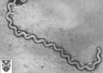
Bacteria are often maligned as the causes of human and animal disease (like this one, Leptospira, which causes serious disease in livestock). However, certain bacteria, the actinomycetes, produce antibiotics such as streptomycin and nocardicin; others live symbiotically in the guts of animals (including humans) or elsewhere in their bodies, or on the roots of certain plants, converting nitrogen into a usable form. Bacteria put the tang in yogurt and the sour in sourdough bread; bacteria help to break down dead organic matter; bacteria make up the base of the food web in many environments. Bacteria are of such immense importance because of their extreme flexibility, capacity for rapid growth and reproduction, and great age - the oldest fossils known, nearly 3.5 billion years old, are fossils of bacteria-like organisms.
Bacteria are microscopic organisms whose single cells have neither a membrane-enclosed nucleus nor other membrane-enclosed organelles like mitochondria and chloroplasts. Another group of microbes, the archaea, meet these criteria but are so different from the bacteria in other ways that they must have had a long, independent evolutionary history since close to the dawn of life. In fact, there is considerable evidence that you are more closely related to the archaea than they are to the bacteria.
Properties of Bacteria
- prokaryotic (no membrane-enclosed nucleus)
- no mitochondria or chloroplasts
- a single chromosome
- a closed circle of double-stranded DNA
- with no associated histones
- If flagella are present, they are made of a single filament of the protein flagellin; there are none of the "9+2" tubulin-containing microtubules of the eukaryotes.
- ribosomes differ in their structure from those of eukaryotes
- have a rigid cell wall made of peptidoglycan.
- The plasma membrane (in Gram-positive bacteria) and both membranes in Gram-negative bacteria are phospholipid bilayers but contain no cholesterol or other steroids.
- no mitosis
- mostly asexual reproduction
- any sexual reproduction very different from that of eukaryotes; no meiosis
- Many bacteria form a single spore when their food supply runs low. Most of the water is removed from the spore and metabolism ceases. Spores are so resistant to adverse conditions of dryness and temperature that they may remain viable even after 50 years of dormancy.

Classification of Bacteria
Until recently classification has done on the basis of such traits as:
- shape
- bacilli: rod-shaped
- cocci: spherical
- spirilla: curved walls
- ability to form spores
- method of energy production (glycolysis for anaerobes, cellular respiration for aerobes)
- nutritional requirements
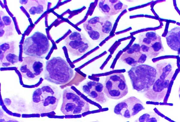
- reaction to the Gram stain.


The Gram stain is named after the 19th century Danish bacteriologist who developed it.
- The bacterial cells are first stained with a purple dye called crystal violet.
- Then the preparation is treated with alcohol or acetone.
- This washes the stain out of Gram-negative cells.
- To see them now requires the use of a counterstain of a different color (e.g., the pink of safranin).
- Bacteria that are not decolorized by the alcohol/acetone wash are Gram-positive.

Protozoa

Protozoa are a diverse group of unicellular eukaryotic organisms, many of which are motile. Originally, protozoa had been defined as unicellular protists with animal-like behavior, e.g., movement. Protozoa were regarded as the partner group of protists to protophyta, which have plant-like behaviour, e.g., photosynthesis.
Protozoa commonly range from 10 to 52 micrometers, but can grow as large as 1 mm, and are seen easily by microscope. The largest protozoa known are the deep-sea dwelling xenophyophores, which can grow up to 20 cm in diameter.
They were considered formerly to be part of the protista family. Protozoa exist throughout aqueous environments and soil, occupying a range of trophic levels.
No hay comentarios:
Publicar un comentario