jueves, 26 de julio de 2012

Peripheral nerves and reflexes


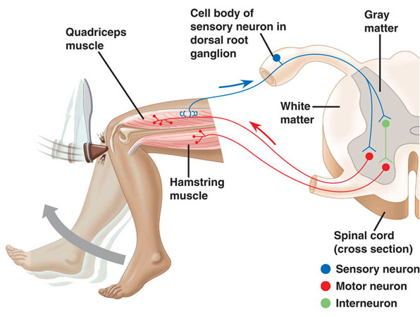
Testing of reflexes.


The peripheral nervous system (PNS) is composed of cranial nerves, spinal nerves, the  distal branches of these nerves, and  ganglia. Any  disorder of nerve  function is  called a neuropathy.  Your textbook describes methods of testing for neuropathies of the cranial nerves. The spinal cord and spinal nerves are tested by  assessing  various somatic reflexes. Testing a reflex helps a clinician evaluate not only the  individual  components of a reflex arc (receptors, neurons,  and muscles) but also the overall state of the nervous  system. Abnormalities of reflex function,  coupled  with other information  gathered  during  the physical examination, provide valuable clues to diagnosis. One advantage of reflex testing is  that it is easy to do; it requires only simple tools and  good powers of observation. Although just a few reflexes are routinely tested, many others can be tested if necessary. Reflexes are usually graded on a scale of 0 to 4+:

0 Areflexia, absence of response
1+ Hyporeflexia, a somewhat diminished response
2+ An average or normal response
3+ Hyperreflexia, a stronger than normal response, possibly indicating disease
4+ Intense hyperreflexia with sustained clonus, indicating disease

Areflexia or hyporeflexia typically indicates a segmental  lesion of the spinal cord segment or nerve root that innervates the  muscle.
Hyperreflexia typically indicates a suprasegmental  lesion of higher levels of the CNS that normally inhibit the reflex. The following discussion describes a few of the deep and superficial reflexes most  commonly tested in adults and some of  the reflexes tested in infants.


Deep Reflexes
The testing of  deep reflexes  usually  involves striking the skin with a  reflex hammer to stretch specific tendons and stimulate the tendon organs and muscle spindles. Clinicians  commonly test the biceps, knee, and ankle reflexes and may  test for clonus.


Biceps Reflex     
The patient  lies supine with the elbow  flexed about 30°. The clinician  presses on the  cubital fossa to  stretch the  biceps  tendon and strikes his or her own fingers with  the  reflex hammer. In a normal response, the  biceps should contract slightly, but not enough to flex the  elbow. If there is a lesion of the  musculocutaneous  nerve or segment C6 of the spinal cord, the  biceps does not contract  but  the finger flexors may  contract slightly. If there is a suprasegmental lesion, the biceps may  contract more forcefully than  normal and the brachioradialis or finger flexors may contract.


Knee  Reflex         
The patient either  sits or  lies supine with the knee flexed 90°. The  examiner strikes the patellar ligament with the  reflex hammer. Normal responses range from a slight twitch of the  quadriceps  femoris muscle t o extension of the knee, lifting the  leg. The  absence of a response indicates a  disease of lumbar  nerve roots L3 and L4 or the femoral nerve.
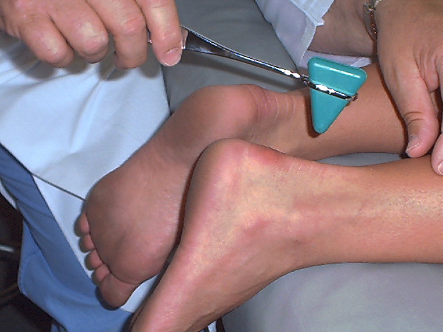
Knee jerk reflex


Ankle Reflex       
There are several  ways to  test this reflex. One is to have the patient kneel on the examining table with the  foot extending beyond the end of the table. The examiner  presses slightly against the foot to dorsiflex it, thus stretching the gastrocnemius muscle, and  then strikes the calcaneal tendon with the reflex hammer. The gastrocnemius should contract and  plantar flex the foot. Other positions  and methods can be tried if this one fails, but if no response is obtained by any method, a disease of  the first sacral nerve  root or the tibial nerve is indicated.


Clonus     
Clonus was  described in  the preceding chapter in connection  with epilepsy  (clonic seizures), but it can also be elicited in  normal persons by the proper test. The patient  should lie supine with the hip and knee flexed at 30° to 45° angles. The examiner then  produces a  sudden and sustained  contraction of the  gastrocnemius and soleus muscles by passively dorsiflexing  the  foot. In normal people, the calf muscles contract, relax, and  contract  again for about two or  three beats. This occurs because the  contraction of  one  muscle stimulates the stretch receptors in the  antagonistic muscle.  When the antagonist contracts, it stimulates the stretch receptors in the original muscle and triggers a reflex  contraction. In people with suprasegmental lesions, the clonus  continues for as long as the examiner dorsiflexes the patient’s foot.


Superficial Reflexes
Superficial reflexes are tested by stimulating the skin. Following are three examples of these tests.

Abdominal Reflex    
The patient  must be supine and relaxed. The examiner strokes the skin of the  abdomen with a pointed object such as a pencil or the handle of a reflex hammer, moving from the lateral margins of the abdomen toward the midsagittal plane along a given dermatome. Normally, the  underlying muscle contracts and pulls the umbilicus toward the stimulus. An absence of response in a  given dermatome may indicate lesions to spinal nerves or roots T7 to T11. The response is often absent, however, in elderly  patients  and people with lax abdominal muscles.


Cremasteric Reflex        
In males, stroking the inner, upper aspect of the thigh with a pin or pencil point  causes the ipsilateral testicle (but not the scrotum) to rise, owing to  contraction of the cremaster  muscle. Lesions in spinal cord segments or nerve roots L1 to L2 or in the corticospinal tracts abolish this reflex.


Plantar Reflex    
To test this reflex, the  patient must be supine with the  lower limbs extended. The examiner strokes the sole of the  foot firmly with the handle point of the reflex hammer, progressing from the heel toward the toes. Normal  subjects show a  flexor plantar response in which  they quickly flex  the hip and knee, dorsiflex the  ankle, and adduct and  plantar flex (curl) the toes. An abnormal extensor plantar (Babinski) response is a reliable, early warning sign of  corticospinal  tract disease;  the patient  extends  and dorsiflexes the great toe and  abducts  (fans) the other toes. The extensor plantar response also sometimes occurs in persons unconscious from  drug or  alcohol intoxication.

Reflexes of Infants
Because  the nervous system is not  completely developed at birth, neurological  examination of infants differs somewhat from the techniques  used for adults.  Normally, a  neonatal examination is performed between 36 and 60 hours after birth. In addition to reflexes, the infant’s motor pattern and body posture are observed. A normal  infant has flexed limbs, and its head may be turned to one side. The  lower limbs  may be moving or kicking, and the  infant is expected to become more  active and to begin crying during the examination. On the other hand, certain  responses are  considered abnormal. For instance, an infant extending its limbs may have suffered  intracranial  hemorrhage. Asymmetric behavior of the upper  limbs suggests brachial plexus palsy.  Lack of increased  activity during the examination  suggests  anoxia or intracerebral hemorrhage.   Infants are tested for the same reflexes asadults as  well as  some additional ones  described here.

The  trunk incurvation (Galant)  reflex is tested by stroking the back from the  shoulder to the buttocks or vice versa, about 1 cm from the midline. This  stimulus should elicit  contraction of  the ipsilateral back  muscles, causing  the  infant’s shoulders and pelvis to curve toward  the  stimulus while  the trunk curves  away. This  response normally disappears at 2 months of  age. Its  earlier absence may indicate a transverse spinal  cord lesion.


The  grasp  reflex is evaluated by  determining the infant’s ability to forcefully grasp the examiner’s hand when the ulnar palmar surface is stimulated. This reflex normally disappears at 3 t o 4 months of age. Persistence of  the reflex beyond 4 months may indicate cerebral dysfunction.


The  rooting  reflex is a response to  tactile stimulation of the lips. When the corner of the baby’s mouth is stroked, the  baby opens its mouth and turns its head toward  that  side.  When the midline of the upper lip is stroked, the  baby extends its head, and when the midline of the lower lip is stroked, the jaw drops. This reflex disappears at 3 to 4 months of  age, although sleeping infants exhibit it at slightly older ages. Absence of  this reflex before 3 to 4 months indicates severe CNS disease.


The  startle  (Moro)  reflex is a response to a sudden stimulus such as a jolt, a loud noise, or  being dropped a short distance (supporting the  baby in a supine  position  and suddenly lowering it about 2 feet). The normal response is for the infant t o extend and abduct all four limbs and extend and fan the  digits,  then flex  and adduct  the  limbs. Neurologic disease is suspected if this reflex persists beyond 4 months and is almost certain if it persists beyond 6  months. An asymmetric response may indicate hemiparesis, brachial plexus injury, or fracture of the clavicle or humerus. The absence of a startle reflex may indicate  kernicterus—damage to the  basal nuclei or  other  areas of the  CNS by accumulated bilirubin, a  hemoglobin breakdown product seen in  hemolytic  disease of  the newborn and some other conditions.


martes, 24 de julio de 2012

Central nervous system.

Poliomyelitis


Poliomyelitis is a viral disease that can affect nerves and can lead to partial or full paralysis.
Causes.

Poliomyelitis is a disease caused by infection with the poliovirus. The virus spreads by:
  • Direct person-to-person contact
  • Contact with infected mucus or phlegm from the nose or mouth
  • Contact with infected feces
The virus enters through the mouth and nose, multiplies in the throat and intestinal tract, and then is absorbed and spread through the blood and lymph system. The time from being infected with the virus to developing symptoms of disease (incubation) ranges from 5 - 35 days (average 7 - 14 days).
Risks include:
  • Lack of immunization against polio
  • Travel to an area that has experienced a polio outbreak
In areas where there is an outbreak, those most likely to get the disease include children, pregnant women, and the elderly. The disease is more common in the summer and fall.
Between 1840 and the 1950s, polio was a worldwide epidemic. Since the development of polio vaccines, the incidence of the disease has been greatly reduced. Polio has been wiped out in a number of countries. There have been very few cases of polio in the Western hemisphere since the late 1970s. Children in the United States are now routinely vaccinated against the disease.
Outbreaks still occur in the developed world, usually in groups of people who have not been vaccinated. Polio often occurs after someone travels to a region where the disease is common. As a result of a massive, global vaccination campaign over the past 20 years, polio exists only in a few countries in Africa and Asia.
Treatment.
The goal of treatment is to control symptoms while the infection runs its course.
People with severe cases may need lifesaving measures, especially breathing help.
Symptoms are treated based on their severity. Treatment may include:
  • Antibiotics for urinary tract infections
  • Medications (such as bethanechol) for urinary retention
  • Moist heat (heating pads, warm towels) to reduce muscle pain and spasms
  • Painkillers to reduce headache, muscle pain, and spasms (narcotics are not usually given because they increase the risk of breathing trouble)
  • Physical therapy, braces or corrective shoes, or orthopedic surgery to help recover muscle strength and function
Prevention.
Polio immunization (vaccine) effectively prevents poliomyelitis in most people (immunization is over 90% effective).


Meningitis.

Meningitis is an inflammation of the membranes (meninges) surrounding your brain and spinal cord, usually due to the spread of an infection. The swelling associated with meningitis often triggers the "hallmark" signs and symptoms of this condition, including headache, fever and a stiff neck in anyone over the age of 2.
Most cases of meningitis are caused by a viral infection, but bacterial and fungal infections also can lead to meningitis. Depending on the cause of the infection, meningitis can resolve on its own in a couple of weeks — or it can be a life-threatening emergency.
If you suspect that you or someone in your family has meningitis, seek medical care right away. Early treatment can prevent serious complications.

Symptoms


It's easy to mistake the early signs and symptoms of meningitis for the flu (influenza). Meningitis signs and symptoms may develop over several hours or over one or two days and, in anyone over the age of 2, typically include:
  • High fever
  • Severe headache that isn't easily confused with other types of headache
  • Stiff neck
  • Vomiting or nausea with headache
  • Confusion or difficulty concentrating — in the very young, this may appear as inability to maintain eye contact
  • Seizures
  • Sleepiness or difficulty waking up
  • Sensitivity to light
  • Lack of interest in drinking and eating
  • Skin rash in some cases, such as in viral or meningococcal meningitis
Signs in newborns
Newborns and infants may not have the classic signs and symptoms of headache and stiff neck. Instead, signs of meningitis in this age group may include:

  • High fever
  • Constant crying
  • Excessive sleepiness or irritability
  • Inactivity or sluggishness
  • Poor feeding
  • A bulge in the soft spot on top of a baby's head (fontanel)
  • Stiffness in a baby's body and neck
  • Seizures
Infants with meningitis may be difficult to comfort, and may even cry harder when picked up.

Causes.


Meningitis usually results from a viral infection, but the cause may also be a bacterial infection. Less commonly, a fungal infection may cause meningitis. Because bacterial infections are the most serious and can be life-threatening, identifying the source of the infection is an important part of developing a treatment plan.
Bacterial meningitis
Acute bacterial meningitis usually occurs when bacteria enter the bloodstream and migrate to the brain and spinal cord. But it can also occur when bacteria directly invade the meninges, as a result of an ear or sinus infection or a skull fracture.

A number of strains of bacteria can cause acute bacterial meningitis. The most common include:
  • Streptococcus pneumoniae (pneumococcus). This bacterium is the most common cause of bacterial meningitis in infants, young children and adults in the United States. It more commonly causes pneumonia or ear or sinus infections.
  • Neisseria meningitidis (meningococcus). This bacterium is another leading cause of bacterial meningitis. Meningococcal meningitis commonly occurs when bacteria from an upper respiratory infection enter your bloodstream. This infection is highly contagious. It affects mainly teenagers and young adults, and may cause local epidemics in college dormitories, boarding schools and military bases.
  • Haemophilus influenzae (haemophilus). Before the 1990s, Haemophilus influenzae type b (Hib) bacterium was the leading cause of bacterial meningitis in children. But new Hib vaccines — available as part of the routine childhood immunization schedule in the United States — have greatly reduced the number of cases of this type of meningitis. When it occurs, it tends to follow an upper respiratory infection, ear infection (otitis media) or sinusitis.
  • Listeria monocytogenes (listeria). These bacteria can be found almost anywhere — in soil, in dust and in foods that have become contaminated. Contaminated foods have included soft cheeses, hot dogs and luncheon meats. Many wild and domestic animals also carry the bacteria. Fortunately, most healthy people exposed to listeria don't become ill, although pregnant women, newborns and older adults tend to be more susceptible. Listeria can cross the placental barrier, and infections in late pregnancy may cause a baby to be stillborn or die shortly after birth. People with weakened immune systems, due to disease or medication effect, are most vulnerable.

Viral meningitis
Each year, viruses cause a greater number of cases of meningitis than do bacteria. Viral meningitis is usually mild and often clears on its own within two weeks. A group of viruses known as enteroviruses are responsible for about 30 percent of viral meningitis cases in the United States. As many viral meningitis episodes never have a specific virus identified as the cause.

The most common signs and symptoms of enteroviral infections are rash, sore throat, diarrhea, joint aches and headache. These viruses tend to circulate in late summer and early fall. Viruses such as herpes simplex virus, La Crosse virus, West Nile virus and others also can cause viral meningitis.
Chronic meningitis
Chronic forms of meningitis occur when slow-growing organisms invade the membranes and fluid surrounding your brain. Although acute meningitis strikes suddenly, chronic meningitis develops over two weeks or more. Nevertheless, the signs and symptoms of chronic meningitis — headaches, fever, vomiting and mental cloudiness — are similar to those of acute meningitis. This type of meningitis is rare.

Fungal meningitis
Fungal meningitis is relatively uncommon and causes chronic meningitis. Occasionally it can mimic acute bacterial meningitis. Cryptococcal meningitis is a common fungal form of the disease that affects people with immune deficiencies, such as AIDS. It's life-threatening if not treated with an antifungal medication.

Treatments and drugs.


The treatment depends on the type of meningitis you or your child has.
Bacterial meningitis 
Acute bacterial meningitis requires prompt treatment with intravenous antibiotics and, more recently, cortisonelike medications, to ensure recovery and reduce the risk of complications. The antibiotic or combination of antibiotics that your doctor may choose depends on the type of bacteria causing the infection. Your doctor may recommend a broad-spectrum antibiotic until he or she can determine the exact cause of the meningitis.

If you or your child has bacterial meningitis, your doctor may also recommend treatments for:
  • Brain swelling
  • Shock
  • Convulsions
  • Dehydration
Infected sinuses or mastoids — the bones behind the outer ear that connect to the middle ear — may need to be drained. Infected fluid that has accumulated between the skull and the membranes that surround the brain also may need to be drained surgically.
Viral meningitis 
Antibiotics can't cure viral meningitis, and most cases improve on their own in a week or two without therapy. Treatment of mild cases of viral meningitis usually includes:

  • Bed rest
  • Plenty of fluids
  • Over-the-counter pain medications to help reduce fever and relieve body aches
If the cause of your meningitis is a herpes virus, there's an antiviral medication available.


Encephalitis

Encephalitis (en-sef-uh-LI-tis) is inflammation of the brain. Viral infections are the most common cause of the condition.
Encephalitis can cause flu-like symptoms, such as a fever or severe headache, as well as confused thinking, seizures, or problems with senses or movement. Many cases of encephalitis may go unnoticed because they result in only mild flu-like symptoms or even no symptoms. Severe cases of encephalitis, while relatively rare, can be life-threatening.
Because the course of any single case of encephalitis is relatively unpredictable, it's important to get a timely diagnosis and treatment.


Nervous tissue.

Guillain barre syndrome.


Guillain-Barre syndrome is a serious disorder that occurs when the body's defense (immune) system mistakenly attacks part of the nervous system. This leads to nerve inflammation that causes muscle weakness.

Guillain Barré syndrome is one of the best examples of a post infectious immune disease and offers insights into the mechanism of tissue damage in other more common autoimmune diseases. Controlled epidemiological studies have linked it to infection with Campylobacter jejuni in addition to other viruses including cytomegalovirus and Epstein Barr virus. The syndrome includes several pathological subtypes, of which the most common is a multifocal demyelinating disorder of the peripheral nerves in close association with macrophages. Evidence from histological examination of peripheral nerve biopsy and postmortem samples suggests that both cell mediated and humoral mechanisms are involved in the pathogenesis. Immunological studies suggest that at least one third of patients have antibodies against nerve gangliosides, which in some cases also react with constituents of the liposaccharide of C jejuni. In the Miller Fisher variant of the disease, these antiganglioside antibodies have been shown to produce neuromuscular block, and may in part explain the clinical signs of that disorder. Treatment with both intravenous immunoglobulin and plasma exchange reduces the time taken for recovery to occur, although mortality remains around 8%, with about 20% of patients remaining disabled.


Antecedent events


Although Guillain, Barré, and Strohl did not comment on the association of this illness with infection, extensive clinical observations supported by epidemiological studies suggest that about 75% of patients have a history of preceding symptoms of infection. Serological studies reveal evidence of antecedent infection in about 30% to 50% of cases. These data are supported by accounts of outbreaks of Guillain Barré syndrome and an association between clinical cases of food poisoning and Guillain Barré syndrome within communities. Case controlled studies confirm a significant association with C jejuni, cytomegalovirus, and probably Epstein-Barr virus. Of these, the association with C jejuni remains the most highly studied. Numerous anecdotal reports of associations with other infections exist in the literature. Some immunisations also appear to be recognised triggers of the disease, including swine flu and rabies. Serological evidence of C jejuni infection occurs in about 30% of patients with Guillain Barré syndrome and appears to be associated with slightly more severe disease and with acute motor axonal neuropathy (AMAN) variants. Many examples of persistent excretion of this organism in the stools of clinical cases of Guillain Barré syndrome are described, strengthening the association.

Pathology


The studies of Asbury and colleagues suggested that the earliest hall mark of Guillain Barré syndrome was the presence of perifascicular lymphocytic cuffs of small vessels in the endoneurium and perineurium. This appears to be associated with demyelination, which is typically macrophage associated. In this regard, the pathology has many similarities with the animal model, experimental allergic neuritis (EAN). More recent pathological studies have shown that several pathological subtypes of Guillain Barré syndrome exist, although the demyelinating form of the disease is the most common, and probably represents at least 75% of cases. Some cases of Guillain Barré syndrome are associated with a primarily axonal process, in which macrophages may be found in close proximity to the axon, with sparing of myelin. This histological finding has been interpreted as indicating an immunological attack on antigens of axonal origin, rather than a myelin antigen in demyelinating forms of the disease.
Still other cases of the disease appear to involve both sensory and motor axons and such cases are termed acute motor and sensory axonal neuropathy (AMSAN). This variant of the disease appears to be the most uncommon and perhaps accounts for only 5% of the clinical syndrome.

Treatment


The mainstay of treatment of Guillain Barré syndrome remains good intensive care, with respiratory support where required and early recognition of respiratory failure. The routine use of prophylaxis for deep venous thrombosis is generally accepted, although it has never been subjected to controlled trial. Positive pressure ventilation with frequent turning to avoid atelectasis and frequent physiotherapy are also useful. Passive limb movement helps to prevent contractures that hinder rehabilitation.
Several specific attempts at treatment have been tried. Unlike the more chronic disorder, chronic inflammatory demyelinating polyneuropathy, Guillain Barré syndrome has not been shown to respond to treatment with oral or intravenous steroids. Several controlled clinical trials have shown that both plasma exchange and intravenous immunoglobulin shorten the time to recovery when used in the early stages of the neuropathy. In the largest study of plasma exchange carried out in North America, this procedure improved the time to achieve walking unaided by 32 days. Whereas plasma exchange clearly removes a blood borne substance mediating the neuropathy, possibly an antibody, the mechanism of action of intravenous immunoglobulin administration is more complicated. This probably includes blockage of Fc receptors, increased catabolism of autoimmune immunoglobulin, and possible roles in providing anti-idiotypic antibodies and in promoting remyelination.

Multiple sclerosis

Multiple sclerosis (MS) is a potentially debilitating disease in which your body's immune system eats away at the protective sheath that covers your nerves. This interferes with the communication between your brain and the rest of your body. Ultimately, this may result in deterioration of the nerves themselves, a process that's not reversible.
Symptoms vary widely, depending on the amount of damage and which nerves are affected. People with severe cases of multiple sclerosis may lose the ability to walk or speak. Multiple sclerosis can be difficult to diagnose early in the course of the disease because symptoms often come and go — sometimes disappearing for months.
There's no cure for multiple sclerosis. However treatments can help treat attacks, modify the course of the disease and treat symptoms.

Symptoms

Signs and symptoms of multiple sclerosis vary widely, depending on the location of affected nerve fibers. Multiple sclerosis signs and symptoms may include:
  • Numbness or weakness in one or more limbs, which typically occurs on one side of your body at a time or the bottom half of your body
  • Partial or complete loss of vision, usually in one eye at a time, often with pain during eye movement (optic neuritis)
  • Double vision or blurring of vision
  • Tingling or pain in parts of your body
  • Electric-shock sensations that occur with certain head movements
  • Tremor, lack of coordination or unsteady gait
  • Fatigue
  • Dizziness
Most people with multiple sclerosis, particularly in the beginning stages of the disease, experience relapses of symptoms, which are followed by periods of complete or partial remission. Signs and symptoms of multiple sclerosis often are triggered or worsened by an increase in body temperature.

Causes

The cause of multiple sclerosis is unknown. It's believed to be an autoimmune disease, in which the body's immune system attacks its own tissues. In multiple sclerosis, this process destroys myelin — the fatty substance that coats and protects nerve fibers in the brain and spinal cord.
Myelin can be compared to the insulation on electrical wires. When myelin is damaged, the messages that travel along that nerve may be slowed or blocked.
Doctors and researchers don't understand why multiple sclerosis develops in some people and not others. A combination of factors, ranging from genetics to childhood infections, may play a role.

Risk factors

These factors may increase your risk of developing multiple sclerosis:
  • Being between the ages of 20 and 40. Multiple sclerosis can occur at any age, but most commonly affects people between these ages.
  • Being female. Women are about twice as likely as men are to develop multiple sclerosis.
  • Having a family history. If one of your parents or siblings has had multiple sclerosis, you have a 1 to 3 percent chance of developing the disease — as compared with the risk in the general population, which is just a tenth of 1 percent. But the experiences of identical twins show that heredity can't be the only factor involved. If multiple sclerosis was determined solely by genetics, identical twins would have identical risks. However, an identical twin has only a 30 percent chance of developing multiple sclerosis if his or her twin already has the disease.
  • Having certain infections. A variety of viruses have been linked to multiple sclerosis. Currently the greatest interest is in the association of multiple sclerosis with Epstein-Barr virus, the virus that causes infectious mononucleosis. How Epstein-Barr virus might result in a higher rate of MS remains to be clarified.
  • Being white. White people, particularly those whose families originated in northern Europe, are at highest risk of developing multiple sclerosis. People of Asian, African or Native American descent have the lowest risk.
  • Living in countries with temperate climes. Multiple sclerosis is far more common in Europe, southern Canada, northern United States, New Zealand and southeastern Australia. The risk seems to increase with latitude.
    A child who moves from a high-risk area to a low-risk area, or vice versa, tends to have the risk level associated with his or her new home area. But if the move occurs after puberty, the young adult usually retains the risk level associated with his or her first home.
  • Having certain other autoimmune diseases. You're very slightly more likely to develop multiple sclerosis if you have thyroid disease, type 1 diabetes or inflammatory bowel disease.

Complications

In some cases, people with multiple sclerosis may also develop:
  • Muscle stiffness or spasms
  • Paralysis, most typically in the legs
  • Problems with bladder, bowel or sexual function
  • Mental changes, such as forgetfulness or difficulties concentrating
  • Depression
  • Epilepsy

Treatments and drugs

There is no cure for multiple sclerosis. Treatment typically focuses on strategies to treat attacks, to modify the course of the disease and to treat symptoms. Some people have such mild symptoms that no treatment is necessary.
Strategies to treat attacks
  • Corticosteroids. The most common treatment for multiple sclerosis, corticosteroids reduce the inflammation that spikes during a relapse. Examples include oral prednisone and intravenous methylprednisolone. Side effects may include increased blood pressure, mood swings and weight gain. Long-term use can lead to cataracts, high blood sugar and increased risk of infections.
  • Plasma exchange (plasmapheresis). This procedure looks a little like dialysis as it mechanically separates your blood cells from your plasma, the liquid part of your blood. Plasma exchange is sometimes used to help combat severe symptoms of multiple sclerosis relapses in people who aren't responding to intravenous steroids.
Strategies to modify the course of the disease
  • Beta interferons. These types of drugs — such as Avonex, Betaseron, Extavia and Rebif — appear to slow the rate at which multiple sclerosis symptoms worsen over time. Interferons can cause side effects, including liver damage, so you'll need blood tests to monitor your liver enzymes.
  • Glatiramer (Copaxone). Doctors believe that glatiramer works by blocking your immune system's attack on myelin. You must inject this drug subcutaneously once daily. Side effects may include flushing and shortness of breath after injection.
  • Fingolimod (Gilenya). An oral medication given once daily, this works by trapping immune cells in lymph nodes. It reduces attacks of MS and short-term disability. To take this drug, you'll need to have your heart rate monitored for six hours after the first dose because the first dose can slow your heartbeat (bradycardia). You'll also need to be immune to the chickenpox virus (varicella-zoster virus). Other side effects include high blood pressure and visual blurring.
  • Natalizumab (Tysabri). This drug is designed to work by interfering with the movement of potentially damaging immune cells from your bloodstream to your brain and spinal cord. Tysabri is generally reserved for people who see no results from or can't tolerate other types of treatments. This is because Tysabri increases the risk of progressive multifocal leukoencephalopathy — a brain infection that is usually fatal.
  • Mitoxantrone (Novantrone). This immunosuppressant drug can be harmful to the heart, and it's associated with development of blood cancers like leukemia, so it's usually used only to treat severe, advanced multiple sclerosis.

Stroke

A stroke occurs when the blood supply to part of your brain is interrupted or severely reduced, depriving brain tissue of oxygen and food. Within minutes, brain cells begin to die.
A stroke is a medical emergency. Prompt treatment is crucial. Early action can minimize brain damage and potential complications.
The good news is that strokes can be treated and prevented, and many fewer Americans die of stroke now than even 15 years ago. Better control of major stroke risk factors — high blood pressure, smoking and high cholesterol — may be responsible for the decline

Symptoms


Watch for these signs and symptoms if you think you or someone else may be having a stroke. Note when your signs and symptoms begin, because the length of time they have been present may guide your treatment decisions.
  • Trouble with walking. You may stumble or experience sudden dizziness, loss of balance or loss of coordination.
  • Trouble with speaking and understanding. You may experience confusion. You may slur your words or have difficulty understanding speech.
  • Paralysis or numbness of the face, arm or leg. You may develop sudden numbness, weakness or paralysis in your face, arm or leg, especially on one side of your body. Try to raise both your arms over your head at the same time. If one arm begins to fall, you may be having a stroke. Similarly, one side of your mouth may droop when you try to smile.
  • Trouble with seeing in one or both eyes. You may suddenly have blurred or blackened vision in one or both eyes, or you may see double.
  • Headache. A sudden, severe headache, which may be accompanied by vomiting, dizziness or altered consciousness, may indicate you're having a stroke.

Causes


A stroke occurs when the blood supply to your brain is interrupted or reduced. This deprives your brain of oxygen and nutrients, which can cause your brain cells to die. A stroke may be caused by a blocked artery (ischemic stroke) or a leaking or burst blood vessel (hemorrhagic stroke). Some people may experience a temporary disruption of blood flow through their brain (transient ischemic attack).
Ischemic stroke
About 85 percent of strokes are ischemic strokes. Ischemic strokes occur when the arteries to your brain become narrowed or blocked, causing severely reduced blood flow (ischemia). The most common ischemic strokes include:

  • Thrombotic stroke. A thrombotic stroke occurs when a blood clot (thrombus) forms in one of the arteries that supply blood to your brain. A clot often may be caused by fatty deposits (plaque) that build up in arteries and cause reduced blood flow (atherosclerosis) or other artery conditions.
  • Embolic stroke. An embolic stroke occurs when a blood clot or other debris forms away from your brain — commonly in your heart — and is swept through your bloodstream to lodge in narrower brain arteries. This type of blood clot is called an embolus.
Hemorrhagic stroke
Hemorrhagic stroke occurs when a blood vessel in your brain leaks or ruptures. Brain hemorrhages can result from many conditions that affect your blood vessels, including uncontrolled high blood pressure (hypertension) and weak spots in your blood vessel walls (aneurysms). A less common cause of hemorrhage is the rupture of an arteriovenous malformation (AVM) — an abnormal tangle of thin-walled blood vessels, present at birth. The types of hemorrhagic stroke include:

  • Intracerebral hemorrhage. In an intracerebral hemorrhage, a blood vessel in the brain bursts and spills into the surrounding brain tissue, damaging brain cells. Brain cells beyond the leak are deprived of blood and damaged. High blood pressure, trauma, vascular malformations, use of blood-thinning medications and other conditions may cause intracerebral hemorrhage.
  • Subarachnoid hemorrhage. In a subarachnoid hemorrhage, an artery on or near the surface of your brain bursts and spills into the space between the surface of your brain and your skull. This bleeding is often signaled by a sudden, severe headache. A subarachnoid hemorrhage is commonly caused by the rupture of an aneurysm, a small sack-shaped or berry-shaped outpouching on an artery in the brain. After the hemorrhage, the blood vessels in your brain may widen and narrow erratically (vasospasm), causing brain cell damage by further limiting blood flow to parts of your brain.
Transient ischemic attack (TIA)
A transient ischemic attack (TIA) — also called a ministroke — is a brief episode of symptoms similar to those you'd have in a stroke. A transient ischemic attack is caused by a temporary decrease in blood supply to part of your brain. TIAs often last less than five minutes.

Like an ischemic stroke, a TIA occurs when a clot or debris blocks blood flow to part of your brain. A TIA doesn't leave lasting symptoms because the blockage is temporary.
Seek emergency care even if your symptoms seem to clear up. If you've had a TIA, it means there's likely a partially blocked or narrowed artery leading to your brain, putting you at a greater risk of a full-blown stroke that could cause permanent damage later. It's not possible to tell if you're having a stroke or a TIA based only on your symptoms. Up to half of people whose symptoms appear to go away actually have had a stroke causing brain damage.

Treatments and drugs

Emergency treatment for stroke depends on whether you're having an ischemic stroke blocking an artery — the most common kind — or a hemorrhagic stroke involving bleeding into the brain.
Ischemic stroke
To treat an ischemic stroke, doctors must quickly restore blood flow to your brain.

Emergency treatment with medications. Therapy with clot-busting drugs (thrombolytics) must start within 4.5 hours if they are given into the vein — and the sooner, the better. Quick treatment not only improves your chances of survival but also may reduce the complications from your stroke. You may be given:
  • Aspirin. Aspirin, an anti-thrombotic drug, is an immediate treatment after an ischemic stroke to reduce the likelihood of having another stroke. Aspirin prevents blood clots from forming. In the emergency room, you may be given a dose of aspirin. The dose may vary, but if you already take a daily aspirin for its blood-thinning effect, you may want to make a note of that on an emergency medical card so doctors will know if you've already taken some aspirin.
    Other blood-thinning drugs, such as heparin, also may be given, but this drug isn't proven to be beneficial in the emergency setting so it's used infrequently. Clopidogrel (Plavix), warfarin (Coumadin), or aspirin in combination with extended release dipyridamole (Aggrenox) may also be used, but these aren't usually used in the emergency room setting.
  • Intravenous injection of tissue plasminogen activator (TPA).Some people who are having an ischemic stroke can benefit from an injection of a recombinant tissue plasminogen activator (TPA), also called alteplase, usually given through a vein in the arm. This potent clot-busting drug needs to be given within 4.5 hours after stroke symptoms begin if it's given into the vein. This drug restores blood flow by dissolving the blood clot causing your stroke, and it may help people who have had strokes recover more fully. Your doctor will consider certain risks, such as potential bleeding in the brain, to determine if TPA is the most appropriate treatment for you.
Emergency procedures. Doctors sometimes treat ischemic strokes with procedures that must be performed as soon as possible.
  • Medications delivered directly to the brain. Doctors may insert a long, thin tube (catheter) through an artery in your groin and thread it to your brain, and then release TPA directly into the area where the stroke is occurring. The time window for this treatment is somewhat longer than for intravenous TPA but still limited.
  • Mechanical clot removal. Doctors may use a catheter to maneuver a tiny device into your brain to physically grab and remove the clot.